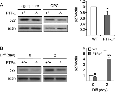
FIGURE 5.
Decreased expression of p27 in PTPα−/− OPCs and differentiating OLs. A, p27 expression was determined by immunoblotting with anti-p27 and anti-actin antibodies, and quantified (graph) for OPCs. B, WT and PTPα−/− mouse oligospheres were dissociated and seeded on PDLO-coated dishes for 2 days in OPC proliferation medium (day 0) followed by incubation for the indicated times in OPC differentiation medium (day 2). The expression of p27 was determined by immunoblotting with anti-p27 and anti-actin antibodies. A and B, band intensity of p27 was normalized to that of actin.

