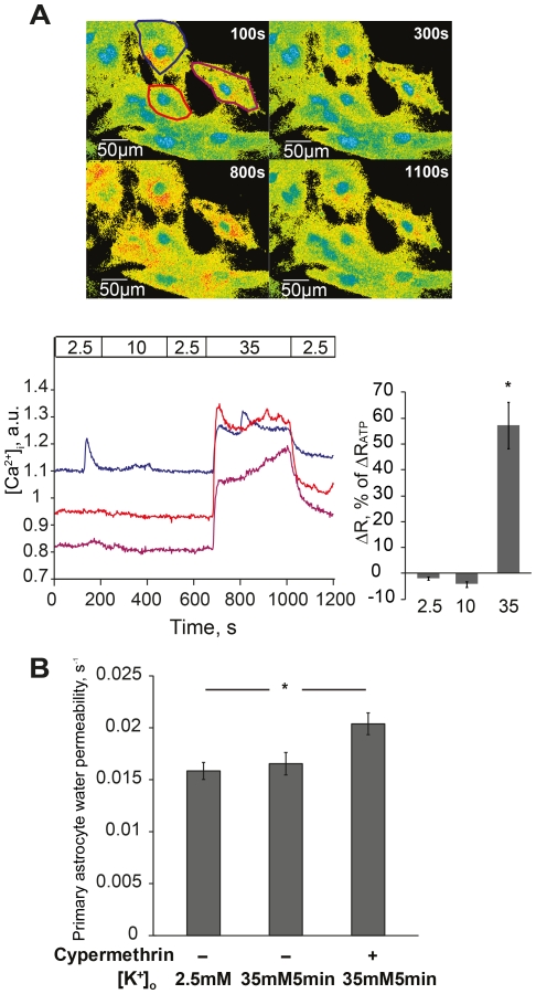
Figure 3. Highly elevated potassium prevents astrocyte water permeability increase via calcium signaling.
(A) Upper panel shows representative 340/380nm fluorescence ratio images of astrocyte primary cultures loaded with Fura-2 AM at indicated time points, respectively. Lower left panel shows representative curves of intracellular calcium ([Ca2+]i) recordings in three individual cells during perfusion with potassium (marked with matched colors in upper panel). Potassium concentrations (mM) are marked in the horizontal bar. Arbitrary units (a.u.) represent ratio values corresponding to [Ca2+]i changes. 10mM potassium (∼200–500s) did not cause any change in [Ca2+]i. 35mM potassium (∼680–1000s) induced a global [Ca2+]i increase. The [Ca2+]i increase disappeared when potassium was returned to baseline (2.5mM). One cell (blue) exhibits a spontaneous [Ca2+]i peak at about 180s. Lower right panel shows summarized calcium data normalized to the peak [Ca2+]i induced by ATP, n = 103. (B) After preincubation with the calcineurin inhibitor cypermethrin, 35mM potassium significantly increased astrocyte water permeability at 5min (*p<0.01, n = 73–89). Values are means±SEM, n; number of cells.

