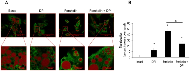
Figure 6. DPI abrogated forskolin-induced HSL translocation from the cytosol to the lipid droplet.
Human differentiated adipocytes were grown and differentiated in 35 mm coated glass bottom dishes and then treated with or without 10 µM DPI in the absence or presence of 5 µM forskolin for 15 min in KRB containing 1% BSA and 0.5 mM oleate. Cells were then fixed with paraformaldehyde and subsequently incubated with anti-total HSL antibody (green), cells were also labeled with Lipotox red for lipid droplet detection (red), as described in the methods section. Cells were imaged in these 35 mm dishes in an inverted microscope. Confocal pictures were taken (A) Images were analyzed using ImageJ software. (B) The panel shows normalized mean values ± S.E.M. for three independent experiments with at least 50 cells per condition per experiment; * p <0.05 vs. basal, # p<0.05 vs. forskolin.

