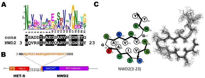
Figure 3. The N-terminal end of the NWD2 protein matches the HET-s PFD consensus.
A. Comparison of the NWD2 sequence with the HET-s consensus sequence. B. Diagram showing the genome organization of het-s and nwd2 as two divergently transcribed adjacent genes. C. Fitting of the NWD2 (3-23) sequence into the β-solenoid fold model (left) and homology model of the NWD2(3-23) sequence (right). Polar residues are given in green, hydrophobic residues are given in white and positively and negatively charged residues in blue and red, respectively.

