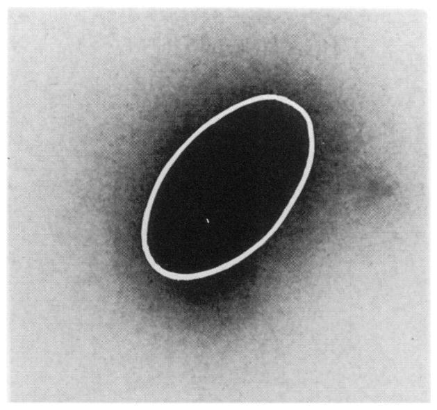
FIG. 2.
Autoradiograph (5× magnification) of a mouse testis injected along the long axis of the organ with a 3-μl standard volume of solution containing 212Pb in equilibrium with its daughter products. The ellipsoidal geometry of the testis (long axis ~7.6 mm, short axis ~5 mm) is outlined by the white line. The low-density halo around the organ is from crossfire exposure due to the finite thickness of the sample (up to 2 mm at the center).

