
Figure 5.
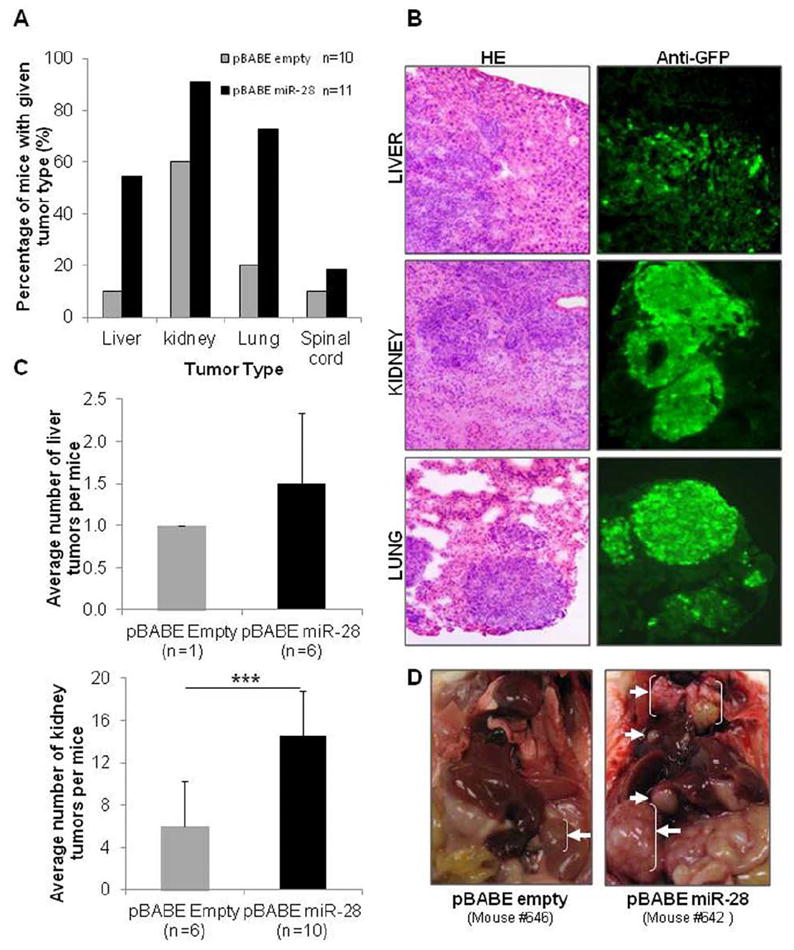
miR-28 increases metastasis in vivo. (A and B) HCT116-pBABE-empty (control) and HCT116-pBABE-miR-28 (stably expressing miR-28) were injected in the vein tail of mice. (A) Thirty-five days postinjection metastases were detected in the liver, kidney, lung, and spinal cord. The percentage of mice with metastases in these organs was consistently higher in miR-28-expressing tumors than in the control. (B) Microscopy images (x100) show hematoxylin and eosin (HE) and anti-green fluorescent protein (GFP) immunohistochemical staining for liver, kidney, and lung metastatic tumors. (C) Number of tumors observed within the liver and kidneys. (D) Photographs of HCT116-pBABE-empty (left panel) and HCT116-pBABE-miR-28 (right panel) mice show the sites with metastasis (white arrows) found in more than 30% of each group of mice.