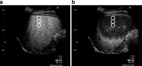
Fig. 2.
Contrast-enhanced images of a 56-year-old woman with chronic hepatitis, in the 5-min phase. a Before instantaneous high-power emission (IHPE): the liver parenchyma showed homogeneous enhancement. Three regions of interests (ROIs, white circles) were placed longitudinally in the centre of the image from 10 to 30 mm in depth. b After IHPE: the liver parenchyma around the liver surface appeared as a hypo-enhancement area because of the breakdown of microbubbles by IHPE. The difference in signal intensity (dB) between the two images was calculated and the average difference in the signal intensities was defined as “intensity difference”

