Figure 2.
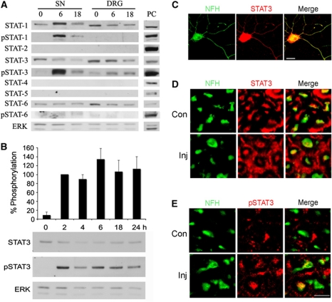
Activation of STAT3 in sciatic nerve axons after injury. (A) Western blot analyses of STAT family members in sciatic nerve (SN) axoplasm or DRG extract from control or injured rat SN (6 or 18 h after injury). Only STAT3 is phosphorylated in both SN and DRG after injury. ERK was used as a loading control. PC denotes positive control (HeLa extract for STATs-1-4 and 6; K562 cells extract for STAT-5; HeLa cells were treated with IFNα for pSTAT-1 and -3; or with IL-4 for pSTAT-6). The experiment was repeated four times with similar results. (B) Western blot analysis of 20 μg aliquots of rat sciatic nerve axoplasm from 0 to 24 h post lesion reveals prolonged phosphorylation of STAT3 after injury. ERK was used as a loading control. Quantification is in percentage of pSTAT3 levels at 2 h post lesion (average±s.e.m.; n=5). (C) Immunostaining for STAT3 and the axonal marker NFH on adult rat DRG neurons in culture shows that STAT3 is found in NFH-positive processes. Scale bar 20 μm. (D) Immunostaining for STAT3 and (E) for pSTAT3 on cross-sections of control or injured (6 h post lesion) rat sciatic nerve reveals the presence of STAT3 in NFH-positive axons in vivo and its phosphorylation after injury. Scale bar 10 μm. Figure source data can be found in Supplementary data.

