Figure 5.
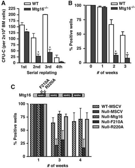
Inactivation of Mtg16 impairs stem cell self-renewal. (A) Graphical representation of the numbers of methylcellulose colonies formed during serial replating assays where 2 × 10⁴ cells were plated every 7 days for 4 weeks (wild type, empty bars; Mtg16-null, full bars). Data are expressed as mean±s.e.m. An unpaired two-tailed t-test indicated these differences were statistically significant (marked by an *; first, P=0.03; second, P=0.01; third, P<0.001; n=4). Representative results from an experiment done in duplicate that are consistent with other biological replicates are shown. (B) Graphical representation of the numbers of methylcellulose colonies formed at the indicated times from LTC-IC cultures of wild-type (open bars) and Mtg16-null (full bars) bone marrow cells. Data are expressed as mean±s.e.m. An unpaired two-tailed t-test indicated the difference at week 2 and week 3 after culture (marked with an *) was statistically significant (P<0.05; n=4). Representative results from three separate experiments are shown. (C) Mtg16 re-expression, but not Mtg16-F210A, complements the LTC-IC defect in Mtg16-null bone marrow cells. Schematic diagram shows the positions of the F210A (blocks Mtg8 binding to HEB and Mtg16 suppression of E protein-dependent transcription) and R220A (control mutation that does not affect E protein binding in the crystal structure of Mtg8) point mutants.

