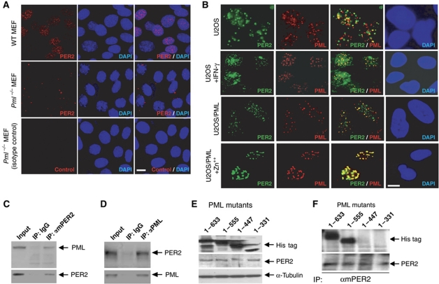
Figure 1.
PML interacts with PER2. (A) Endogenous mPER2 in MEFs: Wild type (WT, top panel) and Pml−/− (middle and lower panel). (B) PML colocalization with PER2. Double IF staining with PER2 and PML antibodies in U2OS cells with (second row) or without (first row) treatment with 1500 U/ml γ-interferon, or stable U2OS/PML cell induced with (fourth row) or without Zn++ (third row). Western analyses were carried out with mPER2, PML or His tag as primary antibody. (C) Western analysis of immunoprecipitates using IgG or mPER2 antibody. (D) Western analysis of immunoprecipitates using IgG or PML antibody. (E) Western analysis of lysates from cells transfected with cDNA's expressing various deletion mutants of PML. (F) Western analysis of immunoprecipitates pulled down from lysates of PML mutants using an mPER2 antibody. Bars: 10 μm.

