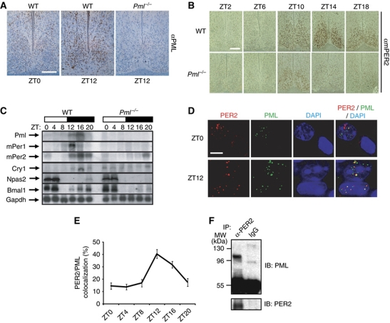
Figure 2.
PML in the SCN and peripheral clocks upregulates mPER2 expression. (A) Immunohistochemical staining with anti-PML monoclonal antibody of SCN obtained from wild-type and Pml−/− mice. Bar: 200 μm. (B) Temporal analysis by immunohistochemical staining with mPER2 antibody of SCN sections obtained from wild-type and Pml−/− mice. Bar: 200 μm. (C) Northern analysis of liver total RNA from wild-type and Pml−/− mice obtained at various ZT points for expression of Pml, mPer1, mPer2, Cry1, Bmal1, Npas2 and Gapdh. Blots were hybridized with radiolabelled cDNA probes for the respective genes. (D) Immunohistochemical staining of PER2 (red) and PML (green) in the wild-type mice SCN at ZT0 and ZT12 observed under × 1000 magnification. Colocalization of PER2 and PML is shown as yellow. Note the nuclear or cytosolic distribution of PER2 at ZT0 in two adjacent nuclei that differentially expressed PML. Bar: 10 μm. (E) Percentage of PER2 and PML in the SCN that colocalized between ZT0 and ZT20 (n>10). (F) Western analysis with anti-PML antibodies of immunoprecipitates from anti-PER2 antibody or IgG with SCN lysates prepared from 20 mouse brains at ZT14.

