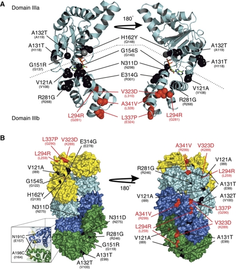
Figure 3.
The DnaA hypermorph and suppressor substitutions are located in domain III. (A) A cartoon representation of monomeric DnaA from the T. maritima crystal structure (PDB ID: 2Z4S) bound to ADP (stick). Domains IIIa and IIIb are separated by a dashed line. The SojG12V hypermorph (black) and suppressor (red) substitutions are shown as spacefill representations. The identity and positions of the B. subtilis amino-acid substitutions are indicated above the corresponding residue of the T. maritima protein. (B) The majority of the hypermorphic substitutions are located either adjacent to, or buried within, the DnaA:DnaA interface. A surface representation of the helical DnaA structure from A. aeolicus (PDB ID: 2HCB) bound to AMP-PCP. DnaA monomers are coloured independently (yellow, cyan, blue, and green). The amino-acid substitutions are coloured and annotated as in (A) above. The inset shows the location of the two residues changed to cysteines for the crosslinking assays, with the black dashed line indicating where BMOE acts.

