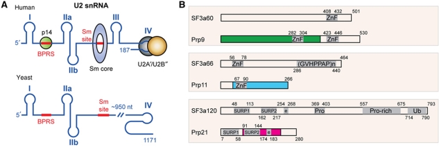
Figure 1.
Structures of U2 snRNA and SF3a subunits. (A) A schematic diagram of human and yeast U2 snRNA. Roman numerals denote RNA stem-loop nomenclature. Sm and the BPRS are highlighted with thick red lines. Human U2 snRNA-binding proteins with known 3D structures, including p14, Sm and the U2A′–U2B″ complexes, are indicated at appropriate positions with colour-filled circles (green: p14; grey and brown: U2A′–U2B″) or a ring (blue: Sm). (B) Domain structure of yeast and human SF3a components. Colour-filled boxes indicate regions involved in the formation of the core domain of the yeast SF3a complex. Known sequence motifs are indicated by a grey-shaded area: ZnF, zinc finger; (GVHPPAP)n, repeats of the heptamer sequence; SURP1 or 2, suppressor-of-white-apricot and prp21 motifs; Pro or Pro-rich, proline-rich regions; Ub, ubiquitin-like domain; e, charged residues. Arabic numbers indicate amino acid numbers.

