Figure 3.
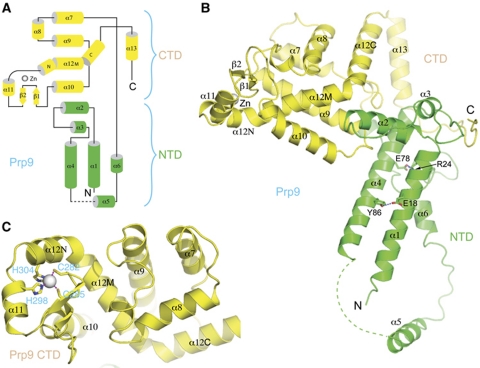
Structure of Prp9ΔC (a.a. 1–389). (A) A topological diagram of the Prp9ΔC fold. Helices are shown as cylinders and strands with arrows. A grey sphere represents the zinc atom. Secondary structural elements shown in green belong to the NTD, and those in yellow form the CTD. (B) A ribbon diagram showing the relative positioning of NTD and CTD. (C) A direct view of the zinc finger in CTD. The two cysteines and two histidines coordinating the zinc ion are shown in a stick representation, and the zinc ion is shown as a sphere. Magenta dashed lines indicate tetrahedral coordination of the zinc ion.

