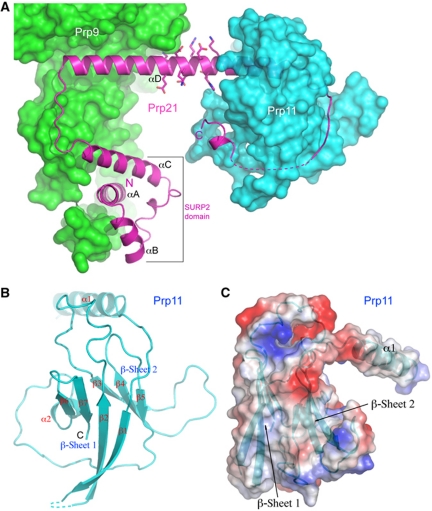
Figure 4.
Structure of Prp21M and Prp11ΔN. (A) Prp21M structure shown as a ribbon diagram (magenta) is superimposed on the surface representations of Prp9ΔC (green) and Prp11ΔN (cyan). A contiguous stretch of charged amino acids on αD is shown as sticks. (B) Structure of Prp11ΔN shown in a ribbon representation. (C) An orthogonal view of Prp11ΔN, shown in an electrostatic potential surface representation, displays a ‘baseball glove’-shaped binding pocket (thumb, α1; palm, β-sheet 2) for Prp21M. A ribbon diagram of Prp11ΔN is superimposed onto the semi-transparent surface.

