Figure 2.
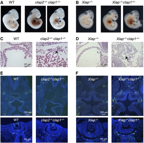
Embryonic lethality of cIap2−/−cIap1−/− and Xiap−/−cIap1−/− DKO embryos. (A, B) Whole view of E10.5 and E11.5 embryos derived from intercrosses of cIap2+/−cIap1+/− and Xiap−/−cIap1+/− mice, respectively. In the DKO embryos, blood has accumulated in the pericardial cavities, but not in those of WT or Xiap−/− littermates. (C, D) Histological analysis of the atria from a WT and cIap2−/−cIap1−/− DKO E10.5 embryo and Xiap−/− and Xiap−/−cIap1−/− DKO E11.5 embryos. The arrow shows discontinuity in the wall. (E, F) TUNEL (green) staining indicating fragmented DNA and nuclear (DAPI, blue) staining of E10.5–E11.5 transverse sections of embryos with genotypes as indicated. Heads are shown in the top panels and eyes are shown in the bottom panels. No gross differences in patterns of TUNEL-stained cells were observed.

