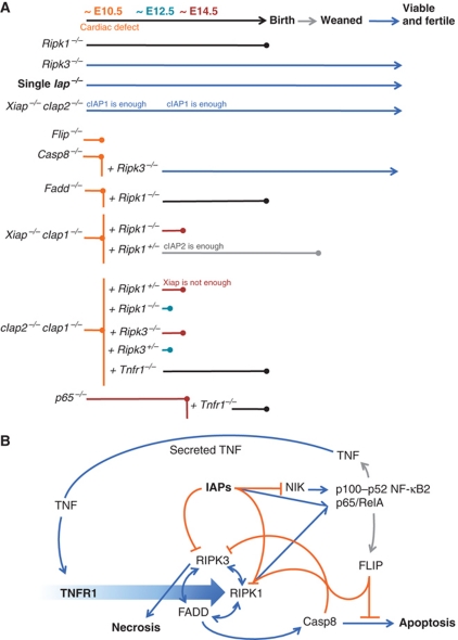
Figure 8.
Roles of cIAPs in signalling and cell death during development. (A) Diagram of extent of viability of single, double, and triple gene deleted mice. (B) Speculative model to account for the phenotypes of gene deleted mice. Binding of TNF to TNFR1 triggers (large blue arrow) formation of complexes that can culminate in cell death by apoptosis or necroptosis, or lead to cell survival. Activating interactions are indicated with blue arrows; inhibitory interactions are indicated with orange lines; transcriptional induction is indicated with grey arrows. Merging of lines (such as those from Casp8 and FLIP to RIPK1, or IAPs and RIPK1 to p65/RelA) indicate proteins that can act together. cIAPs limit levels of NIK, and inhibit cell death mediated by RIPK1 and RIPK3, but cooperate with RIPK1 to activate p65/RelA. RIPK1 has both pro-death and pro-survival functions, by promoting necroptosis via RIPK3, apoptosis via FADD and Casp8, and cell survival via p65/RelA. Casp8 inhibits cell death by necrosis, but promotes cell death by apoptosis. FLIP inhibits cell death by both pathways. This model is not complete, and does not include other important proteins, such as TRADD, TRAF2, CYLD, A20, TAK1, HOL, HOIP, or Sharpin, just to name a few. For further details, see Discussion.

