
Figure 4.
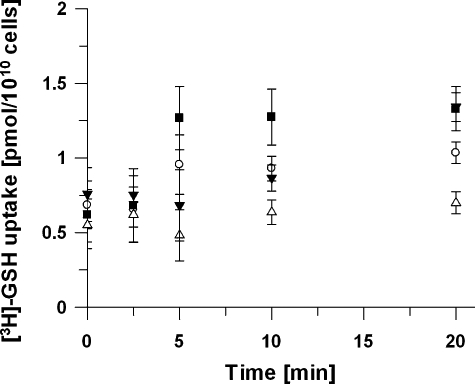
Uptake of [3H]-GSH. Uptake of [3H]-GSH was compared in uninfected (○) and purified trophozoite-infected RBC (▪). Cells were incubated with 24 nM [3H]-GSH and at indicated time points aliquots were withdrawn and the cell-associated label was determined. The amount of [3H]-GSH associated with either uninfected RBC or infected RBC did not significantly increase over 20 min (P > 0.05, unpaired Student t-test). The amount of [3H]-GSH taken up by MACS purified infected RBC after 20 min was not significantly higher than in the control RBC treated in the same way (P > 0.05, unpaired Student's t-test). An excess of unlabelled GSH (3 mM) did not significantly inhibit the amount of [3H]-GSH associated with purified trophozoite infected RBC (▾) but lowered the amount of [3H]-GSH associated with uninfected RBC (Δ) over 20 min. Data represent means of four independent determinations ± SEM.