
Fig. 1.
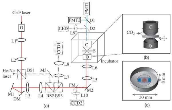
(a) Schematic of the adaptive harmonic generation microscope. Lx, lens; Mx, mirror; BSx, beam splitter; G, Galvo mirrors; DM, deformable mirror; O, objective; S, specimen; C, condenser; Dx, dichroic; PMTx, photomultiplier tubes; FM, flip-mirror. He–Ne laser (dashed outline) is used for DM characterization, and this path is disabled during imaging. Dotted lines show the LED illumination path for wide-field transmitted light imaging. The incubator maintains a stable temperature at 37 °C around the sample stage. (b) A magnified view of the region around the sample. A small plastic chamber around the culture dish, into which a humidified mixture of 5% CO2 in air is supplied to maintain the pH of the culture medium. (c) Illustration of the culture dish showing the placement of the embryos.