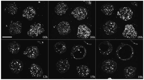
Fig. 5.
Optical sections from time-lapse imaging of mouse embryos showing morula compaction (x and x′) and blastocyst (asterisk) formation. Scale bar is 50 μm. See Video 4 for animated 3-D time-lapse movie and showing a time-lapse sequence of a single optical section. (Color online only.)

