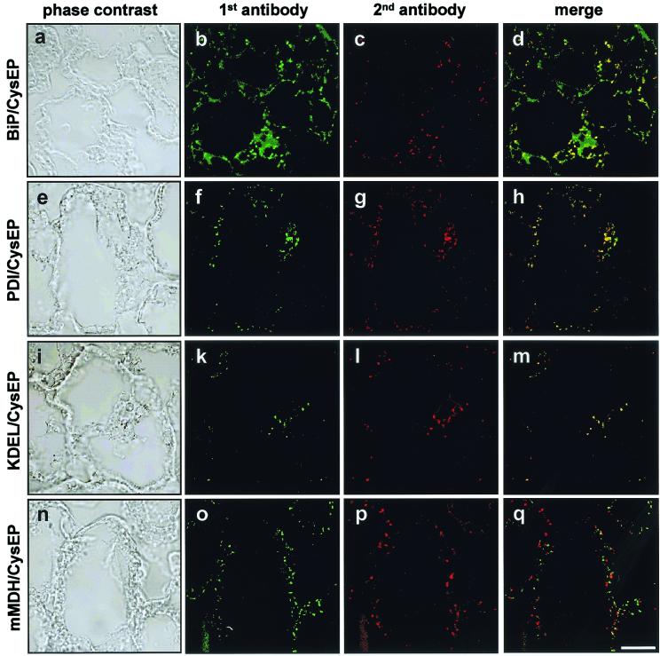
Figure 5.
Double immunofluorescence labeling of 3-day-old germinating castor bean endosperm. (a, e, i, and n) Phase contrast. Sections were first incubated with α-BiP (Hsp70), α-PDI, α-KDEL (MAC256), or α-mMDH (b, f, k, and o; green color), with α-CysEP as the second antibody (c, g, l, and p; red). The merged images (d, h, m, and q) are yellow for BiP, PDI, and KDEL because of colocalization with CysEP, whereas for CysEP and mMDH the red and green dots are separated because of localization in different organelles.

