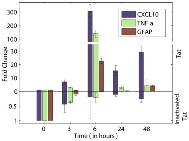
Fig. 2.
Exposure to Tat induces pro-inflammatory factors CXCL10, TNF-α, and GFAP in retinal R28 cells. a CXCL10 and TNF-α showed dramatic rise in R28 cells when incubated with Tat. Expression of these two inflammatory mediators peaked after 6 h of incubation. GFAP expression was also highest after 6 h of incubation. The mRNA expression levels subsequently decreased over the next 48 h. R28 retinal cells were incubated with 200 ng/ml Tat, for up to 48 h, after which the cells were harvested and analyzed for mRNA levels of CXCL10, TNF-α, and GFAP. Data were obtained from four separate experiments, and were analyzed by Students t test. Asterisk indicates statistically significant increases (p<0.05) in factors expressed in cells treated with Tat as compared with vehicle-treated (control) cells. Exposure to inactivated Tat did not induce production of these cytokines or GFAP. Tat was inactivated by boiling for 15 min

