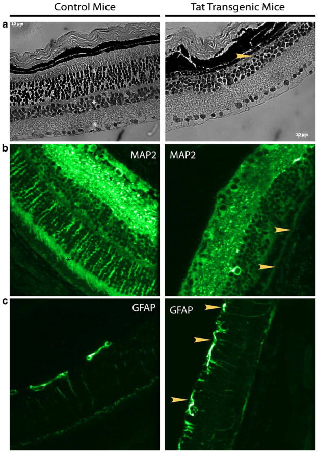
Fig. 4.
HIV-1 Tat-transgenic animals show large scale organizational changes in the retina. Immunocytochemical changes are observed in the retina of HIV-1 Tat-transgenic mice. Representative staining of paraffin-embedded retina sections. Analyses of sections from HIV-1 Tat-transgenic animals show pronounced changes in MAP2 b and GFAP c staining patterns. MAP2 staining is conspicuously missing from the outer plexiform layer and outer photoreceptor segments (arrowhead), of HIV-1 Tat-transgenic animals. GFAP which functions as an activation marker in the retina, showed increased expression with many more processes visible (arrowhead), compared with control animals. Hemotoxylin–eosin staining demonstrated the absence of an entire nuclear layer (arrowhead), absence of the outer photoreceptor segments and thinner retina (a). Scale bar 10 μm. Layers in control animals are marked by asterisks. Eyeballs were harvested from both control animals (n=6) and HIV-1 Tat-transgenic mice. Images shown were captured at ×40 magnification

