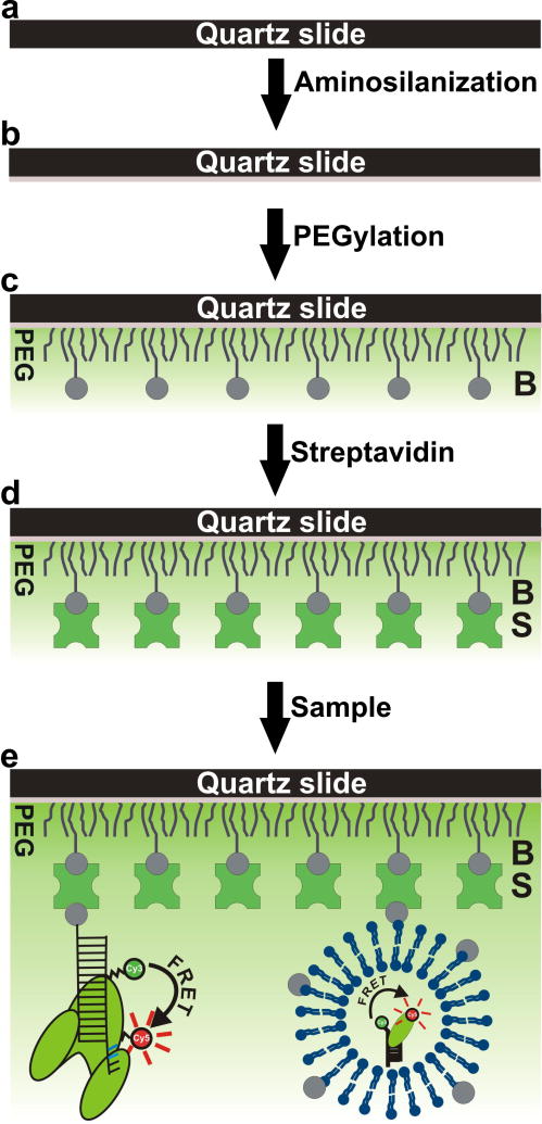
Figure 3.
Stepwise representation of surface passivation and sample immobilization. (a) The slide surface is cleaned. (b) The slide is aminosilanized and ready for PEGylation. (c) PEG and biotin-PEG molecules are conjugated to the amine-modified surface (d) Streptavidin is bound to the immobilized biotin-PEG molecules. (e) The sample can either be surface-tethered with an attached biotin or indirectly immobilized after being encapsulated in a biotinylated lipid vesicle. After washing to remove unbound sample, the slide is ready for use.

