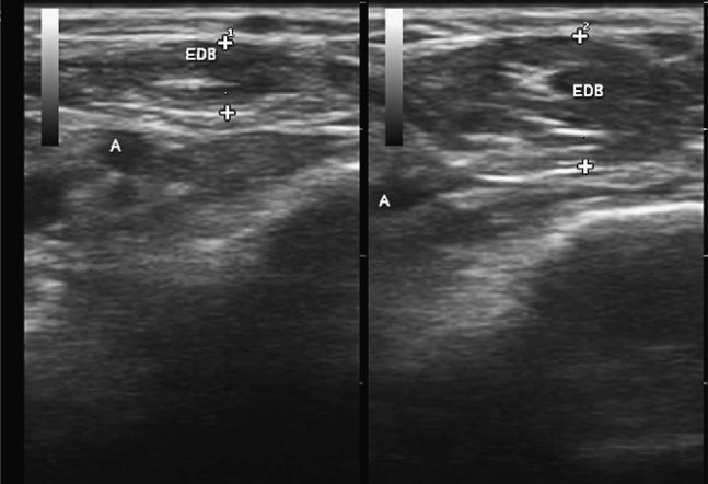
Fig. 1.
Two identical cross-sectional images of the extensor digitorum brevis (EDB). On the left, the muscle (EDB), which is just above a small artery (A), is fully relaxed, and the crosses show the thickness at 2.8 mm. On the right, the muscle is fully contracted, displacing the artery, with a thickness of 5.1 mm.

