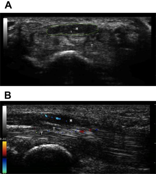
Fig. 6.
(A) Cross-sectional image of the wrist showing a markedly enlarged and somewhat flattened median nerve. The cross-sectional area of the nerve (green tracing) is 31 mm2, which is about 4 times the normal size. Note that the nerve is hypoechoic, particularly when compared with the tendons immediately below it that are much brighter. (B) Sagittal view of the median nerve using color flow Doppler. Note the linear blue elements within the nerve, which demonstrate a stable pulsating blood flow within it, a finding not seen in normal healthy nerves. There is random transient noise in nearby structures such as the tendons (T).

