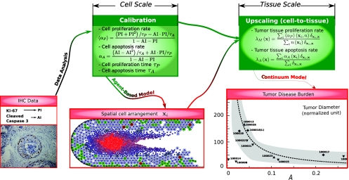
Figure 1.
Flow of information across the formulations at the cellular and tissue scales in a decoupled multiple-scale approach for DCIS.7, 11 Bottom images: (left) Bottom-up calibration: immunohistochemistry measurements from patient tissue used for parameter calibration7 (proliferation and apoptotic indices PI and AI); (center) ABM simulation of DCIS;7 (right) Top-down validation: comparison of model-predicted tumor volume (dashed curve) to the corresponding volume measured from histology of the excised tumors from patients (symbols). Reprinted with permission from Ref. 8. Copyright 2010, Cambridge University Press.

