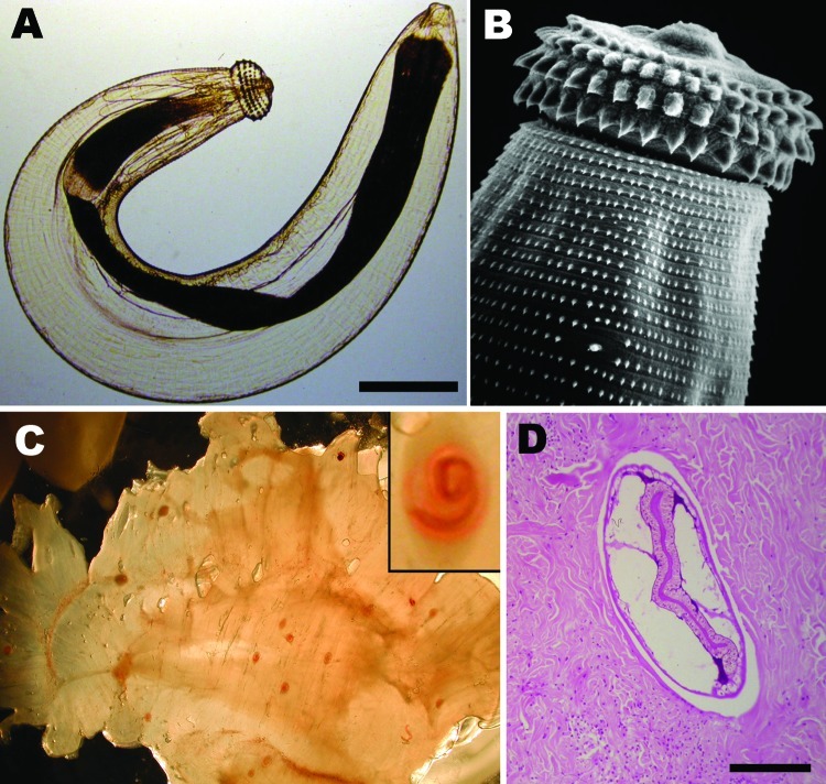
Figure 1.
A) Third-stage larva of the nematode Gnathostoma sp. Scale bar = 250 µm. B) Scanning electronic microscopy image depicting head bulb with 4 cephalic hooklet rows. Original magnification ×500. C) Gnathostoma sp. larvae in the flesh of their intermediate host, Eleotris picta fish. Original magnification ×4. Inset: Higher magnification of an encysted larva; original magnification ×100. Larvae photographs courtesy of Dr Diaz-Camacho, Universidad Autónoma de Sinaloa, Sinaloa, Mexico. D) Cross section of a Gnathostoma sp. larva in human skin biopsy sample (hematoxylin and eosin stain). Scale bar = 250 µm.

