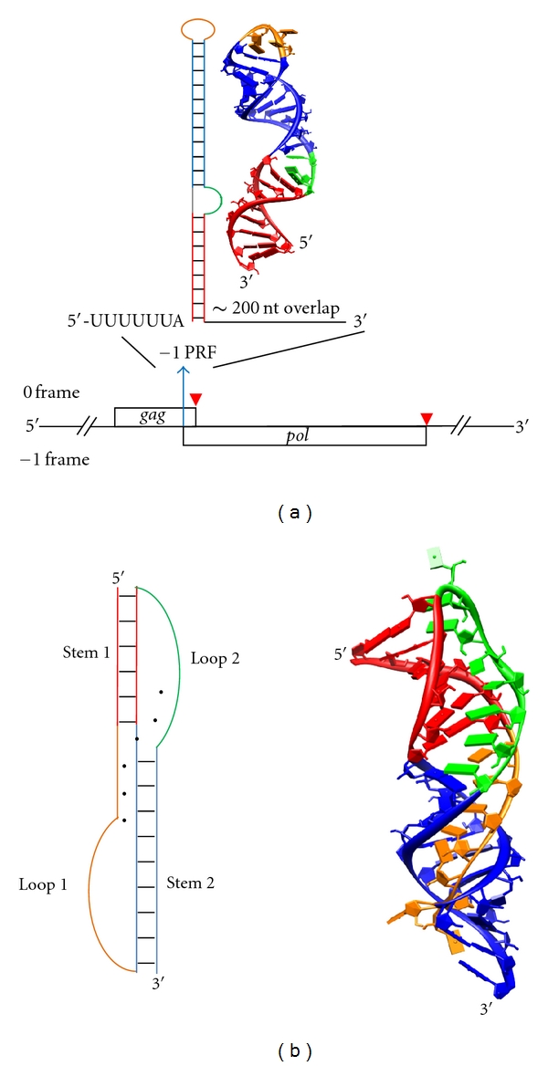
Figure 1.

(a) Structure of HIV gag/gag-pol ORFs, with stop codons indicated by red arrow heads. The pol ORF does not contain a start codon and can only be initiated by −1 PRF within the gag ORF. Therefore, the two ORFs are said to be “overlapping” by a length of ~200 nt [3]. The secondary and NMR-resolved three-dimensional structures for the −1 PRF-stimulating stem-loop are illustrated with corresponding colors, whereas base pairs are indicated by short black bars [20, 40] (PDB 1Z2J). Stems are shown in red and blue, and loops are shown in orange and green. (b) A minimal (ΔU177) human telomerase pseudoknot adapts a canonical H-type pseudoknot configuration [18, 32] (PDB 1YMO). Tertiary major-groove and minor-groove interactions (base triples) are represented by black dots between bases in the secondary structure depiction. The black dot at the junction of Stem 1 and Stem 2 indicates tertiary interaction between Loop 1 (orange) and Loop 2 (green). This RNA structure is not involved in translational regulation, but rather in the activity of the telomerase complex [18, 41]. Its well-known structure makes it an ideal RNA pseudoknot system for studying frameshifting [32, 42]. Stem 1, Loop 1, Stem 2, and Loop 2 are shown in red, orange, blue, and green, respectively. All 3D molecular representations in this paper were produced using the UCSF Chimera package from the Resource for Biocomputing, Visualization, and Informatics at the University of California, San Francisco, USA (supported by NIH P41 RR001081) [43].
