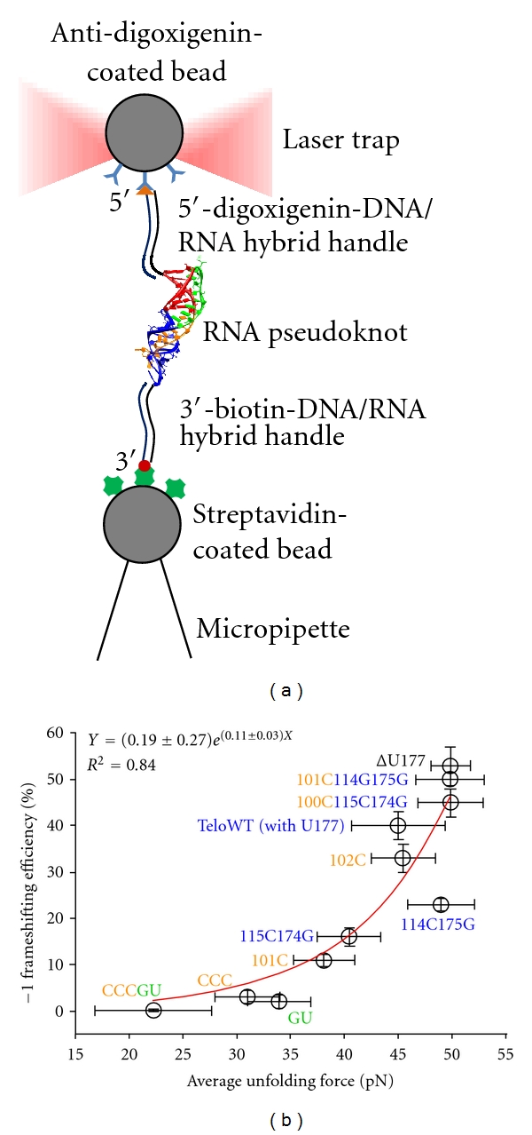
Figure 2.

(a) Schematic representation of optical tweezing. The RNA pseudoknot is flanked by DNA handles that are end-labeled with biotin and digoxigenin, respectively. The handles can then attach to surface-modified beads via biotin-streptavidin and digoxigenin-antibody interactions. Finally, a trapping laser and a micropipette that moves away from the trap produce tension force on the construct. (b) Correlation between −1 PRF efficiencies and externally applied unwinding forces for different pseudoknot constructs. For details of each construct, the reader is referred to reference [32]. Circles indicate average values. Error bars for −1 frameshifting efficiencies along the y-axis are standard errors of the means from in vitro bulk frameshifting assay. Error bars for unfolding forces are standard deviations of the optical tweezing measurements. Frameshifting efficiencies correlate better with unfolding forces (R 2 = 0.84) than melting free energies of pseudoknots (R 2 = 0.59, see [32]). (b) is reproduced with permission from [32].
