Figure 1.
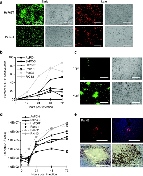
MYXV replicates in pancreatic cancer cells. (a) Pancreatic cancer cells infected with MYXV express early and late markers of viral gene expression. Hs766T or Panc-1 cells were infected with vMyx-GFP at MOI 10 or vMyx-RFP at MOI 5. Fluorescence images and their respective bright field images were taken 24 hours p.i. Bar = 250 µm. (b, c) MYXV spreads in monolayers of pancreatic cancer cells. (b) Cells were infected with vMyx-GFP at MOI 0.1, collected at the indicated time points and analyzed by flow cytometry to determine the percent of infected GFP positive cells. (c) Hs766T cells were infected with vMyx-GFP at MOI 0.1 and analyzed for GFP expression by direct fluorescence 1 day post infection (dpi) and 4 dpi. Bar = 250 µm. (d) MYXV productively infects pancreatic cancer cells. Cells were infected with vMyx-GFP at MOI 0.1, collected at the indicated time points and lysed to determine viral titers. Titers for each sample were performed in triplicate and error bars shown are the mean plus/minus one standard deviation (mean ± SD). (e) MYXV infects pancreatic cancer tumors in vivo. Pan02 (top panels) and Hs766T (lower panels) derived subcutaneous tumors were injected IT with vMyx-tdTr and analyzed for expression of viral reporter genes. Pan02 tumors were excised 3 dpi and analyzed for tdTr expression by direct fluorescence. DAPI was used as a contrast stain. Bar = 200 µm. Hs766T were excised 7 dpi and analyzed for the presence of virus by immunostaining for GFP. Bar = 100 µm. GFP, green fluorescent protein; MOI, multiplicity of infection; MYXV, Myxoma virus; p.i., post infection; RFP, red fluorescent protein; tdTr, tandem dimer tomato red fluorescent protein.

