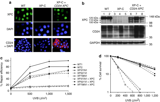
Figure 1.
Restoration of XPC expression, UV-cell survival and nucleotide excision repair in transduced XP-C keratinocytes. (a) Immunofluorescence analysis of XPC (green) and CD24 (red) expression in WT (WT1) and XP-C (XP521VI) keratinocytes before (XP-C) and after (XP-C+CD24-XPC) retroviral transduction and CD24 selection. DAPI was used to stain nuclei. (b) Western blot analysis of WT (1: WT1; 2: WT2), XP-C (3: XP373VI; 4: XP521VI; 5: XP798VI), and transduced XP-C keratinocytes (6: XP373VI+CD24-XPC; 7: XP521VI+CD24-XPC; 8: XP798VI+CD24-XPC). Anti-XPC and anti-CD24 antibodies were used as probes; anti-GAPDH antibody was used as a loading control. (c) Determination of nucleotide excision repair (NER) efficiency after UVB irradiation by unscheduled DNA synthesis (UDS) at the indicated doses. Percent repair efficiency is determined by the ratio of the average number of grains in a strain to the average number of grains in WT#1 cells at 1,500 J/m2 (100%). Data are represented as mean ± SEM of at least two independent experiments. (d) UV survival curves of WT, XP-C, and transduced XP-C strains after UVB irradiation at the indicated doses. GAPDH, glyceraldehyde 3-phosphate dehydrogenase; NER, nucleotide excision repair; UDS, unscheduled DNA synthesis.

