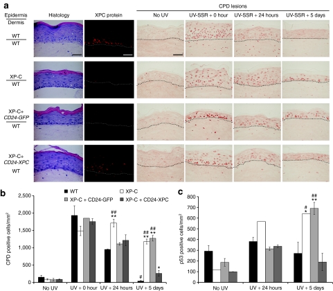
Figure 4.
Restoration of proficient DNA repair in genetically corrected organotypic skin cultures. (a) Histological staining (HES, hematoxylin, eosin, and safran) shows that transduced XP-C (XP798VI) keratinocytes (XP-C+CD24-XPC) regenerate an epidermis with appropriate features of stratification and differentiation. XP-C keratinocytes transduced with pCMMP CD24-IRES-GFP retrovirus were used as a control (XP-C+CD24-GFP). XPC protein expression was revealed by immunofluorescence staining (XPC, red). The NER efficiency after a single UV-SSR exposure (1,600 J/m2 UVB + 17,000 J/m2 UVA) was assessed by indirect immunofluorescence staining of CPD DNA lesions at the indicated time points (CPD lesions, brown). The dash line delimits the dermal–epidermal junction. Bar, 50 µm. (b) Quantification of CPD repair in organotypic skin cultures. For each sample, CPD-positive cells were counted on four independent representative fields and their number was expressed with respect to the epidermis surface area, as estimated using ImageJ software. (c) After p53 immunolabeling, quantification of p53-positive cells was performed as described in b. Mean values obtained from each epidermis were compared to mean values in WT (*) or corrected XP-C epidermis (#) at the same time point. **/##P < 0.001; */#P < 0.01. All XP-C keratinocytes were from patient XP798VI. CPD, cyclobutane pyrimidine dimers; NER, nucleotide excision repair.

