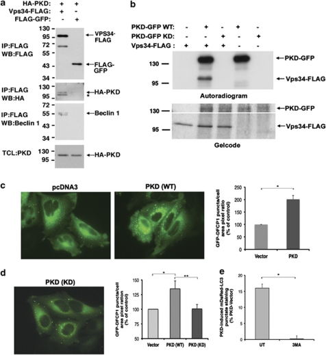
Figure 5.
PKD binds, phosphorylates, and activates Vps34. (a) 293T cells were transfected with HA-PKD and either Vps34-FLAG or FLAG-GFP as control. FLAG-tagged proteins were immunoprecipitated and eluted from the beads. The immunoprecipitated proteins and samples from the total cell lysate (TCL) were separated on SDS-PAGE, transferred to nitrocellulose, and blotted with the indicated antibodies. *Nonspecific band (verified as in control experiments). (b) Immunopurified Vps34-FLAG and either wild-type (WT) or a kinase-dead mutant (KD) of PKD-GFP were quantified against BSA standards and equal protein amounts were incubated in a kinase reaction containing 33P-labeled γ-ATP. (Top) Exposure to X-ray film (autorad); (bottom) gelcode staining (top and bottom parts are different contrast images of the same gel). (c and d) HeLa cells stably expressing GFP-DFCP1 were transfected with pcDNA3 as control and either WT (c) or KD (d) HA-PKD. At 72 h post transfection, the cells were fixed and the ratio of GFP-DFCP1 puncta per total cell area was calculated. Data represent mean+S.D. of three individual experiments. *,**P≤0.05. (e) 293T cells were transfected with mDsRed-LC3 and PKD-GFP and treated with 3MA (5 mM, 16 h) or left untreated (UT). Quantification of cells with punctate GFP-LC3 staining. Data are representative of three individual experiments. *P<0.02

