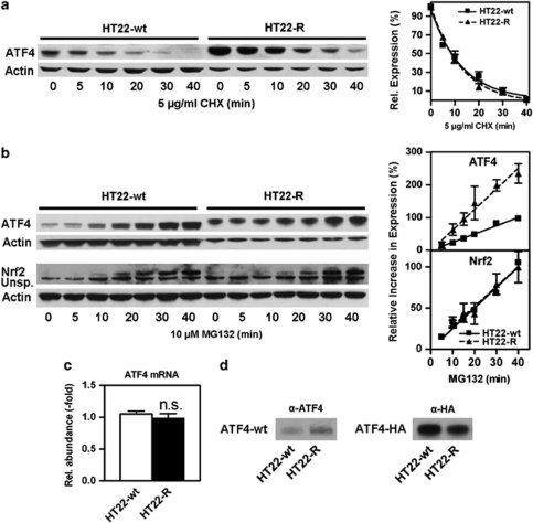
Figure 3.
ATF4 in HT22-R cells has the same half-life and a higher synthesis rate as ATF4 in HT22-wt cells, and the higher molecular weight is not due to post-translational modification. (a) HT22-wt and HT22-R cells were treated with 5 μg/ml cycloheximide (CHX) for the indicated times. Whole-cell extracts were analyzed by western blotting with an antibody against ATF4. Actin served as a loading control. Samples of HT22-R cells were diluted 1 : 3 to adjust for higher ATF4 expression. A representative western blot is shown. The graph represents pooled data from three independent protein preparations with the ATF4/actin ratio without protein synthesis inhibition normalized to 100% as mean±S.E.M. Non-linear regression was performed using one phase exponential decay. (b) HT22-wt and HT22-R cells were treated with 10 μM MG132 for the indicated times and whole-cell extracts were analyzed for ATF4 and Nrf2 expression. For ATF4, HT22-R samples were diluted 1 : 3 to adjust for higher basal ATF4 levels. An unspecific band obtained with the Nrf2 antibody is indicated (Unsp.). The graphs represent three and seven independent experiments for ATF4 and Nrf2, respectively. ATF4 expression was normalized to actin and the relative increase in ATF4 protein was calculated by subtraction of the baseline ATF4/actin ratio at 0 min. ATF4 or Nrf2 to actin ratios obtained in HT22wt cells at 40 min were normalized to 100% for every single experiment and the graphs show the mean±S.E.M. of pooled and normalized data. Statistical analysis was performed by linear regression. (c) qPCR on HT22-wt and HT22-R cDNA shows equal expression of ATF4 mRNA in both the cell lines. The graph represents the mean±S.E.M. of three independent experiments. (d) Recombinant ATF4 does not have a higher molecular weight in HT22-R cells. HT22-wt and HT22-R cells were transfected with wild-type-ATF4 in the pRK7 vector (ATF4-wt) or ATF4-HA. Whole-cell extracts were analyzed by western blotting using antibodies against ATF4 (α-ATF4) or HA (α-HA). For detection of overexpressed ATF4-wt only, samples of HT22-wt and HT22-R cells were diluted 1 : 80 and 1 : 20, respectively, until endogenous ATF4 was undetectable in mock-transfected control cells (not shown). The experiment was done three times with identical results

