Figure 1.
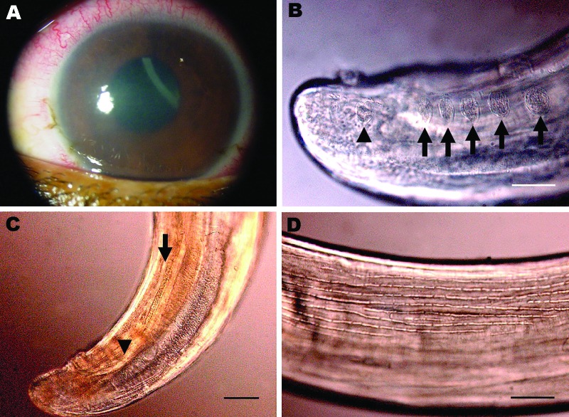
Corneal edema and episcleral hyperemia in the left eye of a 16-year-old boy from Brazil and a free-swimming filarid in the anterior chamber. A) Macroscopic view. B) Five pairs of ovoid pre-cloacal papillae (arrows) and 1 postcloacal caudal papillae (arrowhead). Scale bar = 50 µm. C) Small (arrowhead) and large (arrow) spicules. Scale bar = 40 µm. D) Longitudinal ridges of the area rugosa. Scale bar = 50 µm.

