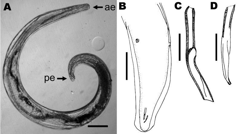
Figure 2.
Parasitic nematode isolated from the eye of the patient, a 29-year-old man from Brazil. A) Nematode that was removed from the iris, showing anterior (ae) and posterior (pe) extremities. Scale bar = 200 µm. B) Caudal region, subdorsal view, showing lateral alae, spicules, and the 2 postdeirids. Scale bar = 150 µm. C) Left spicule; D) right spicule. Scale bars = 20 µm.

