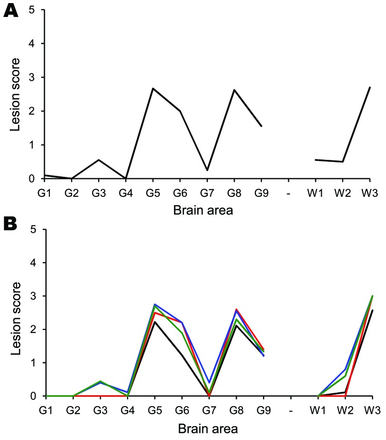
Figure 1.
Tg338 mouse vacuolation lesion profiles of mice infected with scrapie. Only clinically affected mice were considered when generating the lesion profiles. Number in parentheses indicates the mean incubation period of the mice, which contributed to the lesion profile. A) The donor AHQ/AHQ sheep (177 ± 3; n = 10). This profile is compatible with that obtained from other naturally-occurring cases of atypical scrapie (19,20). B) Recipient sheep brain and distal ileum. Cerebellum from animal 12 (179 ± 12; n = 6/10) is indicated in red. Basal ganglion from animal 11 (193 ± 7; n = 10/10) is indicated in blue. Hippocampus from animal 11 (198 ± 11; n = 10/10) is indicated in green. Distal ileum from animal 9 (247 ± 23; n = 9/9) is indicated in black.

