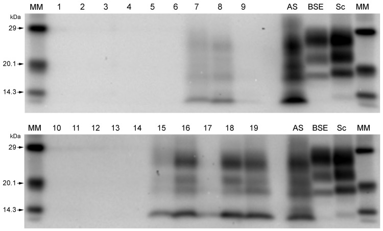
Figure 3.
Western immunoblots showing clear atypical scrapie profiles in sheep in the following brain regions; brainstem of donor ARRa (lane 7), frontal cortex of donor ARRb (lane 8) and frontal cortex of donor AHQ (lane 19). The hippocampus and basal nuclei of recipient animal 11 (lanes 15 and 16, respectively) and cerebellum of recipient animal 12 (lane 18). No discernible signal was seen in the medulla of donor ARRb (lane 9), and only a faint profile was visible for the obex of recipient animal 12 (lane 17). Lane 1, animal 2 obex; lane 2, animal 1 obex; lane 3, animal 3 obex; lane 4, animal 4 obex; lane 5, animal 5 obex; lane 6, animal 6 obex ; lane 7, donor ARRa rostral B.stem; lane 8, donor ARRb frontal cortex; lane 9, donor ARRb caudal medulla; lane 10, animal 7 obex; lane 11, animal 8 obex; lane 12, animal 9 obex; lane 13, animal 10 obex; lane 14, case 11 obex; lane 15, animal 11 hippocampus; lane 1, animal 11 basal nuclei; lane 17, animal 12 obex; lane 18, animal 12 cerebellum; lane 19, donor AHQ frontal cortex; AS, atypical scrapie; BSE, classical bovine spongiform encephalopathy; Sc, classical scrapie; MM, molecular mass marker.

