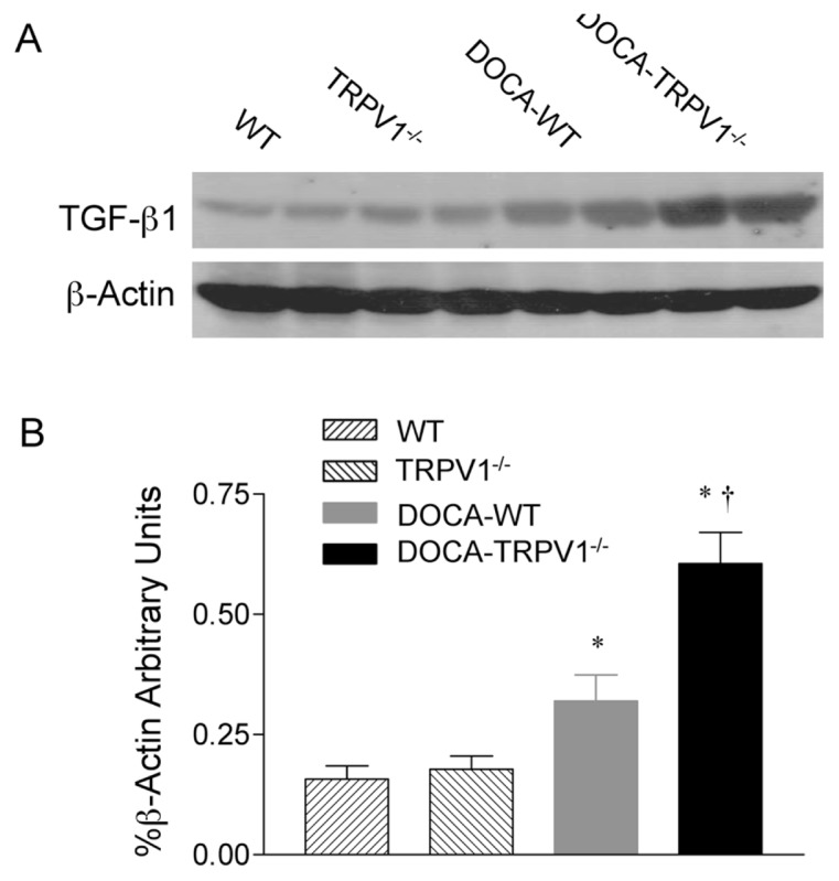
Figure 7.

Changes in renal TGF-β1 protein expression in WT and TRPV1−/− mice after 4 wks of deoxycorticosterone acetate (DOCA)-salt treatment. (A) Representative Western blot of renal TGF-β1 in WT and TRPV1−/− mice with or without DOCA-salt treatment. (B) Bar graph shows the relative optical density values for renal TGF-β1 in WT and TRPV1−/− mice with or without DOCA-salt treatment. Results were expressed as ratio of TGF-β1 to corresponding β-actin. Values are mean ± SE (n = 4 to 5). *P < 0.05 compared to control WT or TRPV1−/− mice; †P < 0.05 compared to DOCA-WT mice.
