
Figure 4.
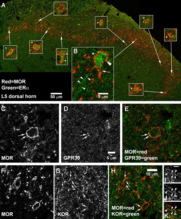
Coexpression of MOR-IR with ERα, GPR30, or KOR in the superficial dorsal horn. A, B, MOR + ERα. A, A montage of the entire superficial dorsal horn of a single section of L5 spinal cord. Insets show 3× higher-magnification views of the cells pointed out by the arrows; cells labeled by ERα are marked with an asterisk (*). ERα-IR was found in 9 of the 10 MOR-IR cells. B, High-magnification view of a cell in the central-lateral superficial dorsal horn. ERα-IR was not restricted to the cell nucleus but also appeared to be in the plasma membrane (large arrowhead) and to extend into proximal dendrites (arrow). ERα-IR was also present in fibrous processes (small arrowheads). The 50 μm scale bar in A applies only to the large montage; magnification of insets is 3× higher. The 5 μm scale bar in B applies only to B. C–E, Expression of GPR30 by an MOR-IR cell in L6 superficial dorsal horn. Arrows mark MOR–GPR30 double labeling. GPR30-IR can also be seen within the cell cytoplasm. F–K, Coexpression of MOR-IR and KOR-IR. F–H, MOR–KOR coexpression in somata. Arrows mark KOR-IR structures visible in MOR-IR neurons in L5 superficial dorsal horn. Double labeling frequently appeared to be in or near the plasma membrane, but sometimes was also seen in the cytoplasm. I–K, MOR–KOR coexpression in fibrous processes. Arrowheads mark a fiber in L5 superficial dorsal horn that was double labeled for MOR and KOR. Scale bar (in I): I–K, is 2 μm.