Figure.
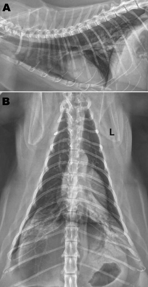
Radiographs of the thorax of a cat with confirmed influenza A pandemic (H1N1) 2009 virus infection. A) Right lateral view; B) dorsoventral view. Asymmetric soft tissue opacities are evident in the right and left caudal lung lobes. An alveolar pattern, composed of air bronchograms with border-effaced (indistinct) adjacent pulmonary vessels, is most pronounced in the left caudal lobe. A small gas lucency in the pleural space appears in the right caudal and dorsal thoracic cavity. An endotracheal tube is visible at the thoracic inlet on the lateral view in this moderately obese cat. L, left.

