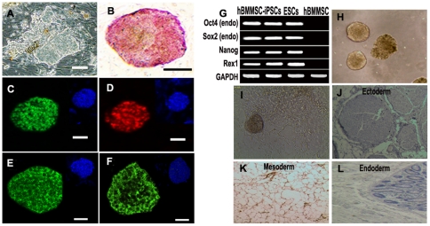
Figure 1. Generation and characterization of iPSCs from hBMMSCs.
Representative hBMMSC-iPSCs colonies (A) and AP staining (B). (C-F) Immunoassay of hBMMSC-iPSC colonies expressing ESC specific markers as OCT4 (C), NANOG (D), TRA-1-81 (E) and SSEA-3 (F). 4, 6-Diamidino-2-phenylindole (DAPI) staining was used to reveal the nuclei. Scale bar, 100 µm. (G) RT-PCR analysis of endogenous pluripotent gene mRNA expression in hBMMSC-iPSCs. Total RNA was isolated from hBMMSC-iPSCs, ESCs and hBMMSCs. (H-L) Pluripotency assay of hBMMSC-iPSCs. EBs was formed (H) and spontaneously differentiated into many types of cells during the in vitro potential assay (I). (J-L) Hematoxylin and Eosin staining of teratoma sections from hBMMSC-iPSCs. 1×10⁷ of hBMMSC-iPSCs were injected subcutaneously into limbs of NOD/SCID mice. Teratomas were obtained after 8–10 weeks. Three germ layer as ectoderm, neural-like tissue (J); mesoderm, adipose tissue (K) and endoderm, intestinal-like epithelium (L) were observed.

