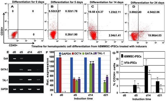
Figure 4. Kinetic expression of the hematopoietic and pluripotent genes during hBMMSC-iPSC commitment to CD34+ progenitor cells, and then to hematopoietic cells.
(A-D) Kinetic expression of CD34+ and CD45+ during the hematopoietic cell differentiation of hBMMSC-iPSCs with flow cytometry analysis. Undifferentiated hBMMSC-iPSCs expressed no CD34 or CD45 (A). After treatment with the cocktail containing mesodermal, hematopoietic, and endothelial inducers for 5 days, hBMMSC-iPSCs expressed about 5% percentage of CD34+ but few CD45+ cells (B). After culturing with the following hematopoietic and endothelial inducer cocktail for additional 7–9 days, the proportion of CD34+ population increased to nearly 20% and a few CD45+ cells were obtained at this stage (C). In hematopoietic potential assay of CD34+ progenitor cells, about 5% population of CD34+CD45+ cells were developed from the CFU assay, and the percentage of CD45+ cells increased to about 25% during the differentiation culture (D). (E-F) Dynamically relative expression of the pluripotent marker Oct4 and the hematopoietic cell markers TAL-1, SCL during hBMMSC-iPSC commitment to CD34+ progenitor cells, and then to hematopoietic cells by RT-PCR (E) and fluorescence intensity (F) assay in the differentiation culture. The values were the mean ± SD of 3 independent experiments. (G) Kinetics of CD34+ cells during hematopoietic differentiation of hBMMSC-iPSCs versus hFib-iPSCs treated with the inducers.

