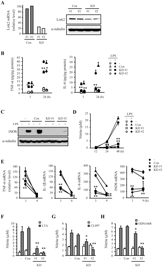
Figure 1. Attenuation of inflammatory responses in Lrrk2-KD microglia.
(A) BV-2 microglia were infected with lentivirus expressing non-targeted (Con) or Lrrk2-targeted shRNA (KD). Two stable clones of each group were selected. Expression levels of Lrrk2 mRNA and protein were analyzed by qRT-PCR (left) and Western blotting (right), respectively. Parental (−), con, and KD cells were treated with or without 100 ng/mL LPS (B–E), 10 µg/mL LTA (F), 500 ng/mL CL097 (G), or 500 ng/ml ODN1668 (H) for indicated times (B, D, E), 12 h (C), 24 h (F), or 48 h (G, H). (B) TNF-α and IL-6 secretion into the culture medium were analyzed by ELISA. (C, D, F–H) iNOS protein expression was assayed by Western blotting (C), and NO release was measured using the Griess reagent, as described in Materials and Methods (D, F–H). (E) TNF-α, IL-1β, IL-6 and iNOS mRNA levels were analyzed by qRT-PCR. Gapdh mRNA and α-tubulin protein levels were analyzed as internal controls for qRT-PCR and Western blotting, respectively. Values are means ± SEMs (*p<0.05, **p<0.01 vs. control). Data are representative of at least three independent experiments unless indicated otherwise.

