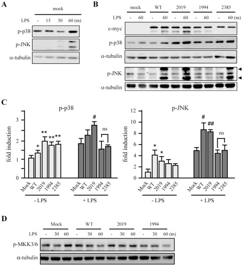
Figure 5. Effect of hLRRK2 overexpression on p38 and JNK phosphorylation in HEK293T cells.
(A) LPS-responsive HEK293T cells (TCM-HEK), prepared as described in Materials and Methods, were co-transfected with empty pcDNA3.1 for the following control experiments. LPS (100 ng/mL) induced phosphorylation of p38, and JNK was as analyzed by Western blotting. (B, D) TCM-HEK cells co-transfected with c-myc-tagged hLRRK2 (WT, G2019S [2019], D1994A [1994], and G2385R [2385]) were treated with LPS (100 ng/mL) for the indicated times. Empty vector (mock) was used as a control. c-myc and α-tubulin were used as markers of LRRK2 expression and loading controls, respectively. Phosphorylation levels of p38, JNK, and MKK3/6 were analyzed by Western blotting. Phosphorylation of JNK was indicated with arrowhead. (C) Band intensities in (B) were quantified using a densitometer. Values are means ± SEMs of three independent experiments (+p = 0.054, *p<0.05, **p<0.01 vs. mock in LPS-untreated group [−LPS]; #p<0.05; ##p<0.01 vs. mock in LPS-treated group [+LPS]; ns, not significant). Data are representative of three independent experiments.

