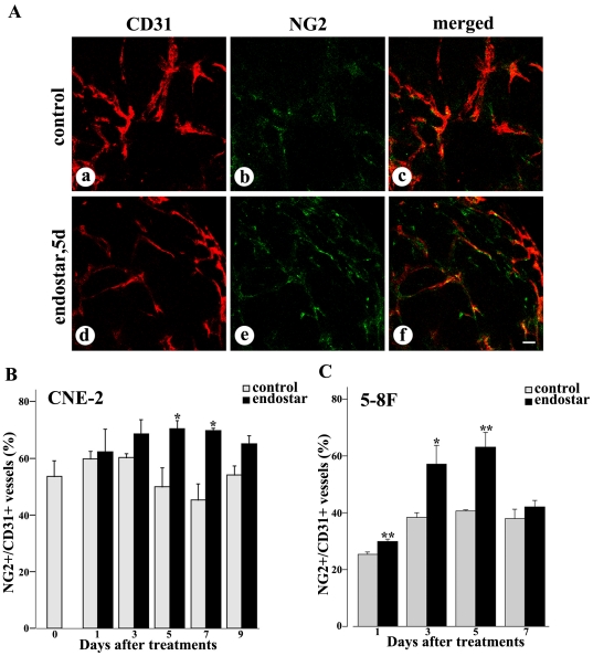
Figure 3. Confocal microscopic images showing comparison of pericyte coverage in NPC tumors with or without endostar treatment.
Fluorescence images of tumors showed CD31-positive endothelial cells (red), NG2-positive pericytes (green), and merged images (orange). A: Some segments of CD31 lacked NG2 immunoreactivity in untreated CNE-2 tumors (a–c). The intensity of NG2 immunofluorescence colocalized with CD31 staining were increased 5 days after endostar treatment in CNE-2 tumors (d–f). B–C: Graphs of the percentage of NG2 showed that pericytes were increased during regression of endothelial cells in both CNE-2 and 5–8F tumors. Quantification of percentages of NG2-positive vessels versus total CD31-positive vessels was determined from 9–12 randomized cryosectioned fields (n = 4–6 mice per group). Columns, means; bars, SEM. *p<0.05 compared with the control group, * *p<0.01 compared with control (Student's t-tests). Scale bar: 50 µm.

