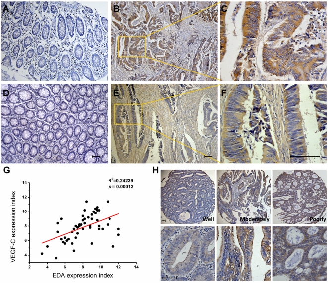
Figure 1. Immunohistochemical staining for EDA and VEGF-C in human colorectal carcinoma.
Representative images of EDA expression in human colorectal carcinoma tissues (B) and in normal colorectal mucosae (A) are shown. Representative images of VEGF-C expression in colorectal carcinoma tissues (E) and in normal mucosae (D) are shown. Inset shows higher magnification of EDA(C) and VEGF-C (F) staining (brown), distinct from the colorectal benign mucosa. (G) Linear regression of EDA and VEGF-C of 52 human colorectal cancer samples was performed. (H) Representative images of EDA expression in tissue microarrays containing tumor samples from 115 CRC patients are shown. The bottom panel shows higher magnification of EDA staining. The slides were counterstained with hematoxylin. Scale bar = 50 µm.

