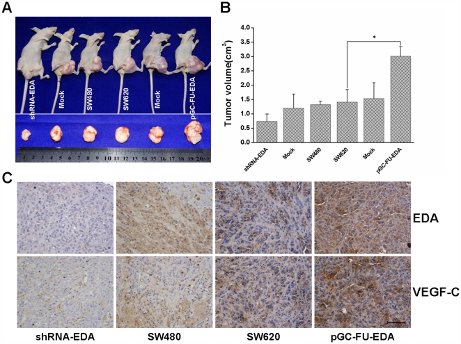
Figure 4. BALB/c nude mice were subcutaneouly injected with transfected cells and negative control cells.
Xenografts were excised and sized 42 days later. (A) Effect of EDA on tumor proliferation. (B) Tumor volumes were measured when mice were sacrificed and the data are presented as mean determinants (±SEM). * p < 0.05. (C) Immunohistochemical staining of EDA and VEGF-C was performed in nude mouse xenografts. Scale bar = 50 µm.

