
Figure 6.
bmp1a Is Expressed in Osteoblasts, which Appear Normally Differentiated in frilly fins
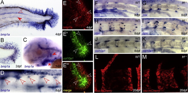
(A–D) In situ hybridization of bmp1a at 4 dpf (A), 2 dpf (B), 3 dpf (C) and 8 dpf (D). Expression is seen in fin mesenchyme cells of the fin fold (A and B), floor plate and hypochord (A, open and filled arrowheads, respectively), branchial arches (C, asterisk), and operculum (C, arrowhead) and perichordal cells of the anterior notochord (D, arrowheads).
(E–E″) Confocal images of double-fluorescent in situ hybridizations showing coexpression of bmp1a (E and E″; red) and the osteoblast marker collagen10a1 (E′ and E″; green) in osteoblasts on the operculum at 4 dpf. Most cells express both markers (three cases highlighted by arrowheads), and central, more mature osteoblasts express slightly higher levels of col10a1.
(F–K) Perichordal expression of osteoblast markers is not disrupted in frf mutants. Lateral images of anterior notochord of 8 dpf WT (F, H, and J) and frf−/− (G, I, and K) larvae hybridized with probes for sp7 (F and G), osteopontin (H and I), and collagen10a1 (J and K).
(L and M) Confocal images of sp7:mCherry-expressing osteoblasts on vertebrae of WT (L) and frf mutant (M) larvae at 20 dpf. There is no reduction in osteoblast numbers in the mutant.